Showing posts sorted by relevance for query Anemia. Sort by date Show all posts

Wednesday, October 6, 2010

HEMOGLOBIN (Hb)

Hemoglobin adalah molekul protein dalam sel darah merah yang membawa oksigen dari paru-paru ke jaringan tubuh dan karbon dioksida dari jaringan ke paru-paru.
  • Hemoglobin terdiri dari empat molekul protein (globulin rantai) yang terhubung bersama-sama.
  • Hemoglobin dewasa normal (Hbg) molekul mengandung rantai 2-globulin alfa dan 2 rantai beta-globulin.
  • Pada janin dan bayi, hanya ada beberapa rantai beta dan molekul hemoglobin terdiri dari 2 rantai alfa dan 2 rantai gamma.
  • Saat bayi tumbuh, rantai gamma secara bertahap diganti dengan rantai beta.
  • Setiap rantai globulin berisi struktur pusat penting yang disebut molekul heme.
  • Tertanam dalam molekul heme adalah besi yang mengangkut oksigen dan karbon dioksida dalam darah
  • Besi yang terkandung dalam hemoglobin juga bertanggung jawab untuk warna merah darah.

  • Hemoglobin juga memainkan peran penting dalam mempertahankan bentuk sel darah merah. struktur hemoglobin abnormal bisa mengganggu bentuk sel darah merah dan menghambat fungsi dan mengalir melalui pembuluh darah.
  • Hemoglobin biasanya diukur sebagai bagian dari jumlah darah lengkap (CBC) dari sampel darah.
Tingkat hemoglobin dinyatakan sebagai jumlah hemoglobin dalam gram (gm) per desiliter (dl) darah utuh, 1 desiliter = 100 mililiter.

Kisaran normal hemoglobin tergantung pada usia dan, mulai pada masa remaja, jenis kelamin orang tersebut. Kisaran normal adalah:

  • Bayi yang baru lahir: 17-22 gm / dl
  • Satu (1) minggu usia: 15-20 gm / dl
  • Satu (1) bulan usia: 11-15gm/dl
  • Anak-anak: 11-13 gm / dl
  • Dewasa laki-laki: 14-18 gm / dl
  • Dewasa wanita: 12-16 gm / dl
Manfaat pemeriksaan Hb:
  1. Pemeriksaaan penyaring utk tegakkan diagnosa.
  2. Pencerminan reaksi tubuh terhadap penyakit
  3. Petunjuk kemajuan terapi.
Kadar Hb normal bervariasi tergantung :
  1. Umur
  2. Jenis Kelamin
  3. Geografi ( tinggi rendahnya daerah ).
Kadar Hb menurun pada ANEMIA dan dapat dijumpai pada :
  1. Thalasemia
  2. Haemoglobinopathy
  3. Perdarahan akut atau kronis
  • Beberapa metode yang ada untuk mengukur hemoglobin, yang kebanyakan dilakukan oleh mesin otomatis saat ini dirancang untuk melakukan beberapa tes darah yang berbeda.
  • Dalam mesin, sel-sel darah merah dipecah untuk mendapatkan hemoglobin menjadi solusi.
  • Hemoglobin bebas adalah yang terkena suatu bahan kimia yang mengandung sianida yang mengikat erat molekul hemoglobin untuk membentuk Cyanmethemoglobin.
Jadi hal yang mendasar dari uraian diatas adalah Hb sangat penting dalam keberlangsungan keberadaan erythrocyt , meskipun selain itu Fe ( zat besi) namun sering kesalahan pemahaman kasus Anemia disebabkan karena kurang zat besi .
  • Kebanyakan orang memahami bahwa anemia disebabkan oleh kekurangan zat besi dalam aliran darah tetapi dalam kenyataannya anemia ditandai oleh defisiensi dalam hemoglobin sel darah merah mengurangi kemampuan darah untuk mengangkut oksigen ke sel-sel kita dan untuk menghilangkan karbon dioksida.
Jenis anemia yang ada di tubuh yang karena gangguan Hb dengan berbagai penyebab .

  1. Thalassmia
  2. Anemia Aplastic
  3. Anemia Bersel Sabit (siklemia)
  4. Anemia Defisiensi Besi
  5. Anemia pernisiosa
  6. Anemia kronis
  7. Anemia hemolitik
Thalessemia

Thalassemia Minor dan Mayor

Thalassemia minor

Ini adalah anemia yang paling umum pada orang dari keturunan Mediterania. Ini adalah kasus anemia ringan. Sel darah lebih kecil dari biasanya sel-sel darah dan mati dengan mudah menyebabkan tekanan untuk reproduksi sel darah merah yang baru karena tidak cukup sel-sel darah baru untuk menggantikan hilangnya sel darah merah.

Thalassmia mayor

Thalassmia utama juga disebut Cooley's anemia. Ini adalah warisan yang mempengaruhi gangguan produksi hemoglobin normal. Thalassmia utama adalah penyakit mematikan tanpa transfusi darah sering, orang-orang seperti ini tidak mungkin bertahan hidup karena besi terbentuk dalam hati dan organ lainnya, menyebabkan gagal jantung.

Anemia Aplastic

Ini adalah salah satu yang paling mematikan dan kasus paling langka . Untuk alasan apapun, sumsum tulang yang menghasilkan sel darah merah tidak bekerja dengan benar atau mungkin disebabkan oleh gangguan auto-imun menghasilkan sel darah putih menyerang sumsum tulang.

Anemia Bersel Sabit

Ini adalah bentuk, serius yang mengancam hidup orang yang mewarisi anemia yang sebagian besar mempengaruhi orang-orang keturunan Afrika. Anemia sel sabit terbentuk dari defisiensi hemoglobin menyebabkan sel darah merah menjadi lengket, kaku, dan lebih rapuh menghasilkan bentuk kurva dan bentuk sabit, membatasi peredaran darah di tubuh kita.

Anemia Defisiensi Besi

Besi adalah mineral yang paling penting dalam darah, bekerja sama dengan seng dan tembaga untuk membantu untuk meningkatkan tingkat oksigen dan sirkulasi darah kita untuk memelihara sel-sel tubuh kita dan mengeluarkan karbon dioksida dari aliran darah. Besi sulit untuk diserap mineral, tanpa bersama-sama dengan vitamin C, zat besi yang paling dalam tubuh kita terbuang.

Anemia pernisiosa ( Defisiensi asam folat dan vitamin B12 anemia)

Asam Folat dan vitamin B12 sangat penting untuk reproduksi sel darah merah yang berlebihan yang disebabkan oleh minum alkohol dan asupan tertentu tertentu seperti obat kontrasepsi oral atau obat antikanker. Orang-orang yang hanya makan makanan dimasak memiliki risiko tertinggi kekurangan folat vitamin B12 dan anemia.

Anemia kronis (Kehilangan darah)

Jenis anemia dihasilkan dari berbagai kondisi kronis seperti wasir, kanker, menstruasi dan tukak lambung.

Anemia hemolitik

Dalam hal ini, penghancuran sel darah merah tua melebihi produksi yang baru yang disebabkan oleh sintesis hemoglobin cacat atau trauma dalam arteri, atau disebabkan oleh antibodi yang diproduksi oleh sistem kekebalan tubuh merusak sel-sel darah merah.


Wednesday, October 18, 2017

SOAL ESSAY DARAH


ESSAY


1. What are the main functions of blood?

  • Blood is a means of substance transportation throughout the body. 
  • Blood distributes nutrients, oxygen, hormones, antibodies and cells specialized in defense to tissues and collects waste such as nitrogenous wastes and carbon dioxide from them.
2. What elements make up blood?
  • Blood is made of a liquid and a cellular portion. 
  • The fluid part is called plasma and contains several substances, including proteins, lipids, carbohydrates and mineral salts. 
  • The cellular components of blood are also known as blood corpuscles and they include erythrocytes (red blood cells), leukocytes and platelets.
3. What is hematopoiesis?
  • Hematopoiesis is the formation of blood cells and the other elements that make up blood.
4. Where does hematopoiesis occur?
  • Hematopoiesis occurs in the bone marrow (mainly within flat bones), where erythrocytes, leukocytes and platelets are made; and in lymphoid tissue, which is responsible for the maturation of leukocytes and which is found in the thymus, spleen and lymph nodes.
5. In which bones can bone marrow chiefly be found? Is bone marrow made of bone tissue?
  • Bone marrow can mainly be found in the internal cavities of flat bones, such as vertebrae, the ribs, the shoulder blades, the sternum and the hips. 
  • Bone marrow is not made of bone tissue, although it is a connective tissue just like bone tissue.
6. What are blood stem cells?
  • Stem cells are undifferentiated cells able to differentiate into other types of specialized cells. 
  • The stem cells of the bone marrow produce differentiated blood cells. 
  • Depending on stimuli from specific growth factors, stem cells are turned into red blood cells, leukocytes and megakaryocytes (the cells that form platelets). 
  • Research shows that the stem cells of the bone marrow can also differentiate into muscle, nervous and hepatic cells.
7. What are the other names for erythrocytes? What is the function of these cells?
  • Erythrocytes are also known as red blood cells (RBCs) or red corpuscles.
  • Red blood cells are responsible for transporting oxygen from the lungs to tissues.
8. What is the name of the molecule in red blood cells that transports oxygen?
  • The respiratory pigment of red blood cells is called hemoglobin.
9. What is the molecular composition of hemoglobin? Does the functionality of hemoglobin as a protein depend on its tertiary or quaternary structure?
  • Hemoglobin is a molecule made of four polypeptide chains, each bound to an iron-containing molecular group called a heme group. Therefore, the molecule contains four polypeptide chains and four heme groups. 
  • As a protein composed of polypeptide chains, the functionality of hemoglobin depends upon the integrity of its quaternary structure.
10. On average, what is the lifespan of a red blood cell? Where are they destroyed? Where do heme groups go after the destruction of hemoglobin molecules?
  • On average, red blood cells live for around 120 days. The spleen is the main organ where old red blood cells are destroyed. 
  • During the destruction of red blood cells, the heme groups turn into bilirubin and this substance is then captured by the liver and later excreted to the bowels as a part of bile.
11. What are the functions of the spleen? Why can people still live after a total splenectomy (surgical removal of the spleen)?
  • The spleen has many functions: it participates in the destruction of old red blood cells; in it specialized leukocytes are matured; it helps regenerate the hematopoietic tissue of bone marrow when necessary; and it can act as a sponge-like organ to retain or release blood into circulation. 
  • It is not impossible to live after a total splenectomy because none of the functions of the spleen are both vital and exclusive to this organ.
12. What is anemia? What are the four main types of anemia?
  • Anemia is a low concentration of hemoglobin in the blood. 
  • The four main types of anemia are nutrient-deficiency anemia, anemia caused by blood loss, hemolytic anemia and aplastic anemia.
  • Nutrient-deficiency anemia is caused by a dietary deficiency in fundamental nutrients necessary for the production or functioning of red blood cells, such as iron (iron deficiency anemia), vitamin B12 and folic acid.
     
  • Anemia caused by blood loss occurs in hemorrhagic conditions or in diseases such as peptic ulcerations and hookworm disease.
     
  • Hemolytic anemia is caused by the excessive destruction of red blood cells, for example, in diseases such as malaria or in hypervolemic conditions (excessive water in blood causing lysis of red blood cells).
     
  • Aplastic anemia occurs from deficiencies in hematopoiesis and occurs when bone marrow is injured by cancer from other tissues (metastasis), by autoimmune diseases, by drug intoxication (such as sulfa drugs and anticonvulsants) or by chemical substances (such as benzene, insecticides, paints, herbicides and solvents in general). Some genetic diseases also affect bone marrow, causing aplastic anemia.
13. What is the difference between white and red blood cells? What are leukocytes?
  • Red blood cells are called erythrocytes and white blood cells are called leukocytes. 
  • Leukocytes are cells specialized in the defense of the body against foreign agents and are part of the immune system.
14. What are the different types of leukocytes and how are they classified into granulocytes and agranulocytes?
  • The types of leukocytes are lymphocytes, monocytes, neutrophils, eosinophils and basophils. 
  • Granulocytes are those with a cytoplasm that contains granules (when viewed under electron microscopy): neutrophils, eosinophils and basophils are granulocytes. 
  • Agranulocytes are the other leukocytes: lymphocytes and monocytes.
15. What is the generic function of leukocytes? What are leukocytosis and leukopenia?
  • The generic function of leukocytes is to participate in the defense of the body against foreign agents that penetrate it or are produced inside the body. 
  • Leukocytosis and leukopenia are clinical conditions in which a blood sample contains an abnormal count of leukocytes. 
  • When the leukocyte count in a blood sample is above the normal level for the individual, it is called leukocytosis. 
  • When the leukocyte count is lower than the expected normal level, it is called leukopenia. 
  • The multiplication of these defense cells, leukocytosis, generally takes place when the body is suffering from infections or in cancer of these cells. A low count of these defense cells, or leukopenia, occurs when some diseases, such as AIDS, attack the cells or when immunosuppressive drugs are used.
     
  • In general, the body uses leukocytosis as a defense reaction when it is facing infectious or pathogenic agents. 
  • The clinical condition of leukocytosis is therefore a sign of infection. 
  • Leukopenia occurs when there is a deficiency in the production (for example, in bone marrow diseases) or excessive destruction of leukocytes (for example, in the case of HIV infection).
16. What are the mechanisms to contain hemorrhage called? 
  • The physiological mechanisms to contain hemorrhage (one of them is blood clotting) are generically called hemostasis, or hemostatic processes.
17. How are platelets formed? What is the function of platelets? What are the clinical consequences of the condition known as thrombocytopenia?
  • Platelets, also known as thrombocytes, are fragments of large bone marrow cells called megakaryocytes. 
  • Through their properties of aggregation and adhesiveness, they are directly involved in blood clotting as well as release substances that activate other hemostatic processes. 
  • Thrombocytopenia is a clinical condition in which the blood platelet count of an individual is lower than normal. In this situation, the person becomes susceptible to hemorrhages.
18. How does the body know that the coagulation process must begin?
  • When tissue wound contains injury to a blood vessel, the platelets and endothelial cells of the wall of the damaged vessel release substances (platelet factors and tissue factors, respectively) that trigger the clotting process.
19. How can the blood coagulation (clotting) process be described?
  • Blood clotting encompasses a sequence of chemical reactions whose products are enzymes that catalyze the subsequent reactions (that is why clotting reactions are called cascade reactions). 
  • In the plasma, thromboplastinogen transforms into thromboplastin, a reaction triggered by tissue and platelet factors released after injury to a blood vessel. 
  • Along with calcium ions, thromboplastin then catalyzes the transformation of prothrombin into thrombin. 
  • Thrombin then catalyzes a reaction that produces fibrin from fibrinogen. Fibrin, as an insoluble substance, forms a network that traps red blood cells and platelets, thus forming the blood clot and containing the hemorrhage.
20. What are clotting factors?
  • Clotting factors are substances (enzymes, coenzymes, reagents) necessary for the clotting process to happen. 
  • In addition to the triggering factors and reagents already described (tissue and platelet factors, thromplastinogen, prothrombin, fibrinogen, calcium ions), other substances participate in the blood clotting process as clotting factors. 
  • One of these is factor VIII, the deficiency of w
21. In what organ are most of the clotting factors produced? What is the role of vitamin K in blood coagulation?
  • Most clotting factors are produced in the liver. 
  • Vitamin K participates in the activation of several clotting factors and is essential for the proper functioning of blood coagulation.
22. What is factor VIII? What is the genetic disease in which this factor is absent?
  • Factor VIII has the function of activating factor X, which in turn is necessary for the transformation of prothrombin into thrombin during the clotting cascade. 
  • Hemophilia A is the X-linked genetic disease in which the individual does not produce factor VIII and as a result is more susceptible to severe hemorrhages.
23. How is hemophilia treated? Why is hemophilia rare in females?
  • Hemophilia is medically treated with the administration of factor VIII, in the case of hemophilia A, or of factor IX, in the case of hemophilia B, by means of blood or fresh frozen plasma transfusions. 
  • Both hemophilia A or B are X-linked recessive diseases. 
  • For a girl to be hemophilic, it is necessary for both of her X chromosomes to be affected whereas boys, who have only one X chromosome, are more easily affected. 
  • A girl with only one affected chromosome does not present the disease, since the normal gene of the unaffected X chromosome produces the clotting factor.
24. What is the epidemiological association between hemophilia and HIV infection?
  • Since hemophilic patients need frequent transfusions of clotting factors (VIII or IX) they are more susceptible to contamination by infectious agents present in the blood from which the transfused elements come. In the past, blood banks did not usually perform HIV detection tests and many hemophilic patients have become infected with the virus.
25. What are anticoagulants? What are the practical applications of anticoagulants, such as heparin, in Medicine?
  • Anticoagulants are substances that block clotting reactions and therefore stop the coagulation process. 
  • Ordinarily, anticoagulants circulate in the plasma, since under normal conditions blood must be maintained fluid. 
  • In Medicine, anticoagulants such as heparin are used in surgeries in which tissue injuries caused by surgery act could trigger undesirable systemic blood clotting. 
  • They are also used to avoid the formation of thrombi inside the blood vessels of patients riskat an increased risk for thrombosis.
26. What is dicoumarol? What is the role of this substance in the clotting process and what are some examples of its toxicity?
  • Dicoumarol is an anticoagulant drug. Due to its molecular structure, dicoumarol competes with vitamin K to bind to substrates, thus blocking the formation of clotting factors and interrupting the production of prothrombin. 
  • Dicoumarol is found in some decomposing vegetables and can cause severe internal hemorrhages when those vegetables are accidentally ingested. 
  • Coumarinic anticoagulants cannot be administered during pregnancy since they pass the placental barrier and can cause fetal hemorrhages.
27. Streptokinase is a substance used in the treatment of acute myocardial infarction. What is function of this substance?
  • Substances known as fibrinolytics, such as streptokinase and urokinase, can destroy thrombi (clots formed inside blood vessels, capillaries or within the chambers of the heart) and are used in the treatment of obstructions of the coronary arteries or other blood vessels. 
  • Streptokinase destroys the fibrin network and as a result dissolves the thrombotic clot. Its name is derived from the bacteria that produce it, streptococci.

Wednesday, April 27, 2011

HASIL CHECK DARAH

Habis ini kamu harus cek darah sebelumnya puasa ya , nanti hasil cek itu bawa ke saya sehingga kami bisa mendapatlan analisa kamu sakit atau normal.OK ( sekilas dialog seorang pasien dan dokter )

Ada 8 point dalam pelaksanan chek darah secara sederhana '

  1. Hb ( Hemoglobin) ……….g/dl
  2. Haematocrite ( Hct )
  3. Laju endap darah (ESR)……….mm/jam
  4. Jumlah Sel Darah Putih ………..x10³/mm³
  5. Hitung Jenis Sel Darah Putih ( Diff Counting)
  6. Jumlah Sel Darah Merah…………. Jt/mL
  7. Jumlah trombosit………………/mm³
  8. Indeks eritrosit.

Manfaat pemeriksaan darah lengkap :
  1. Sebagai Pemeriksaaan penyaring untuk membantu diagnosa.
  2. Sebagai Pencerminan reaksi tubuh terhadap suatu penyakit.
  3. Dapat dipakai sebagai petunjuk kemajuan penderita anemia atau infeksi.


HAEMOGLOBIN ( Hb ) :

Haemoglobin berfungsi mengangkut oksigen ke jaringan. Molekul haemoglobin tersusun dari haem dan globin. Haem terbentuk dari Fe dan protoporphyrin yang terbentuk di mito
Kondria. Globin terbentuk dari rantai asam amino dalam ribosom.
Daya ikat Hb terhadap O2 menurun : mudah melepaskan O2 terjadi dalam keadaan :
  • bila kadar 2,3 –DPG menurun
  • kadar H+ atau CO2 meningkat.

Nilai normal Hb ( bervariasi ) :
  • Laki-laki : 13,4 – 17,7 g/dl
  • Wanita : 11,4 – 15,1 g/dl
  • Neonatus : 16,5 + 3 g/dl
  • Anak : 3 bln : 12,0 + 1,5 g /dl

Manfaat pemeriksaan Hb:
  1. Pemeriksaaan penyaring utk tegakkan diagnosa.
  2. Pencerminan reaksi tubuh terhadap penyakit
  3. Petunjuk kemajuan terapi.


Kadar Hb normal bervariasi tergantung :
  1. Umur
  2. Jenis Kelamin
  3. Geografi ( tinggi rendahnya daerah ).
Kadar Hb menurun pada ANEMIA dan dapat dijumpai pada :
  1. Thalasemia
  2. Haemoglobinopathy
  3. Perdarahan akut atau kronis

Pada Infeksi Kronik :
  • Lactoferin : transferin likiron – binding protein
  • Ambil Fe dari transferin yang beredar
  • Komposisi dengan transferin sewaktu ambil Fe dari macrophage

Lekemia :
  • Fisiologis : Hamil karena proses hemodilusi RBC↓ Hb ↓
  • Hb : Policetemia : Jumlah RBC ↑
  • Dehidrasi :RBC↑+ Hb↑karena cairan tubuh banyak yang hilang


PEMERIKSAAM KADAR Hb

Metode KALORIMETRI

1. Direct Matching
  • Warna drh dibandingkan dengan warna standar.
  • Cepat, sederhana, menyenangkan , namun Kesalahannya besar, tidak tepat

2. Alkali Hematin
  • Darah + Na oH dididihkan Hbà hijau biru dari larutan, alkali hematin à Standar / Spectrophotometer
  • Akurat
  • Tidak akurat untuk ukur Hb bayi

3. Metode Oxyhemoglobine
  • Darah + Na2 Co3 / NH4OH à Oxyhemoglobin à Spectropht
  • Cepat, akurat
  • Oxyhemoglobin + Cu à methemoglob shg hasil lebih rendah

4. Metode cyanmethemoglobine
  • Darah ( Hb ) + lar Drabkin K3Fe(CH)6à MetHb
  • MetHb + KCN à CyanmetHb diperiksa dengan Spectrophotometer 540 nm dibandingkan dengan standard.
  • Cepat, teliti kecuali Sulhemoglobine
  • Mengandung CN yg bersifat racun

5. Metode Asam Hametin ( Sahli )
  • Hb direaksikan dg Hcl à asam hematin (sempurna) à diencerkan
  • Dibaca pada skala tabung sahli sesuaikan dengan standard
  • Cepat, sederhana, tidak mahal
  • Kurang teliti, kesalahan + 5 s/d 10 %


HEMATOKRIT ( HCT ) = PCV ( Packed Cell Volume )

Prosentase volume sel darah merah thd vol darah seluruhnya
( Darah + anticoagulan à dipusingkan )
Normal : Dewasa Laki : 45 – 47 %, Dewasa Wnt : 40 – 42 %
Hematocrit meningkat pada :
  • Peningkatan Juml RBC : Policitemia
  • Penurunan vol plasma
  • Makrositosis

o Hematocrit menurun pada :
  • Anemi
  • Micrositosis
  • Dilusi = hidrasi

Metode Px. Hct :
  • Makro = Wintrobe
  • Micro = Tabung kapiler
  • Elektronik = Auto Analysa, Caulter Caunter

Penyebab kesalahan pemeriksaan :
1. Sample darah diambil setelah terjadi perdarahan ( Hematocrit cenderung tinggi )
2. Anticoalugan berlebih
3. Kecepatan & waktu pemusingan ( Macro 30’, Mikro 5-10’ )
4. Terlalu lama Vena terbendung


LAJU ENDAP DARAH ( LED )
= ESR ( erytrocyt sedimentation rate )
  1. Kecepatan RBC mengendap setelah memisahkan diri dari plasma
  2. Ukuran : mm/jam
  3. Menggambarkan komposisi plasma dan perbandingan antara eritrocit & plasma
  4. Setiap keadaan yg meningkatkan penggumpalan sel satu dgn yg lain akan meningkatkan LED.

Tahapan :
  1. Terbentuknya Rouleaux
  2. Vase pengendapan cepat
  3. Vase pengendapan lambat

Faktor-faktor yang mempengaruhi :
  1. Faktor sel darah merah ( massa yg terbentuk stlh rouleaux ) Bentuk tertentu sel darah merah Aglutinasi Makrosit RBC yg rendah
  2. Plasma : Alfa globulin Alfa2 globulin Fibrinogen

3. Faktor mekanis dan teknis
  • Posisi tabung LED yg panjang & diameter tabung sterilitas
  • Sterilitas
  • Suhu
  • Kondisi darah ( Antikoagulan, darah simpan lama ).

Cara Pemeriksaan :
  1. Makro ( 1 s/d 2 ml darah ) : Westergren, Wintrobe, Culter
  2. Mikro ( 1 tetes darah ) : Landau, Hellinger, Cresta.

Harga Normal :
Laki-laki Wanita
  • Westergren 0 – 15 mm/jam 0 – 20 mm/jam
  • Wintrobe 0 – 10 mm/jam 0 – 20 mm/jam
  • FK Unair 2 – 13 mm/jam 2 – 12 mm/jam

HITUNG LEKOSIT ( WBC = WHITE BLOOD CELL )

  • Dengan kamar penghitung IMPROVED NEUBAUER

Harga Normal : ± 4 – 10 x 109/ dl / cmm
  • Laki : 4,7 – 10,3 x 10⁹/l
  • Wanita : 4,3 – 11,3 x 10⁹ /l

Variasi jumlah sel darah putih :
  1. Jumlah yg masuk peredaran darah dipengaruhi oleh bakteri, endotoksin, besar pori dinding sinusoid, tingkat maturasi sel.
  2. Jumlah yg keluar dari peredaran darah
  3. Distribusinya
  4. Kombinasi 1 s/d 3

Faktor-faktor yg mempengaruhi keseimbangan Netrophil :
  1. Latihan fisik ( Epinephrin )
  2. Endotoksin
  3. Kortikosteroid

Pemeriksaan Mikroskopis :
  • Manual
  • Kamar hitung Neubauer
  • Hemositometer

Alat yang dipakai :
  • Mikroskop
  • Pipet Lekosit
  • Kamar hitung
  • Larutan pengencer Leukosit ( Turk, asam aeetat )

Pemeriksaan Automatic : Elektronik

HITUNG JENIS SEL DARAH PUTIH

  • Menghitung dan mengelompokan WBC yg tampak dihapusan darah dari 100 – 200 sel
  • Berperan dalam diagnosa penyakit
  • Normal ada 6 jenis WBC matur :
  • Eo / Ba / Neu stab / Neu seg / Limfosit / Mo


ABNORMALITAS

  1. Penyimpangan prosentase jenis WBC Peningkatan Eo : alergi, cacing Ba : CML, Policitemia Vera, dll
  2. Sel plasma : measles, varicella, MM
  3. Limfosit abnormal : paling sering Mononukleosis infeksiosa
  4. Sel darah putih muda
  5. Dewasa : Mieloblas, promieloblas, mielosit à AML, CML Anak : Limfosit à ALL


HITUNG ERITROSIT ( RBC = RED BLOOD CELL )

Pengukuran jumlah RBC.
  • Saat lahir jumlah RBC paling tinggi, berangsur turun saat Dewasa.
  • RBC dibentuk dalam sumsum tulang pipih & proximal dari tulang panjang.
  • Umur RBC 120 hari dalam peredaran darah.
Harga NORMAL :
  • Laki 2 dws : 4,3 jt – 5,9 jt/mL
  • Wanita dws : 3,9 jt – 4,8 jt/mL
  • Bayi : 5,0 jt – 7.0 jt/mL
  • Anak 3 bl : 3,2 jt – 4,8 jt/mL
  • Anak 1 th : 3,6 jt – 5,2 jt/mL
  • Anak 10-12 th : 4,0 jt – 5,4 jt/mL
Untuk penghitungan jumlah RBC dapat dipakai :
  • Manual : Kamar Hitung Improved Neubauer setelah diencerkan dgn larutan Hayem.
  • Elektrik

HITUNG TROMBOSIT ( PLT = PLATELET )
  • Pada penderita dgn riwayat perdarahan atau purpura, monitoring pada pemberian obat yang potensial atau diperkirakan beracun pada sumsum tulang, monitoring terapi heparin, monitoring setelah splenektomià jumlah trombosit harus dimonitor.
Jumlah NORMAL TROMBOSIT : 150.000 -400.000 /mm³

  • Perdarahan spontan terjadi pada Plt < 20.000/mm³ terjadi
  • Pada : Penurunan fs sumsum tulang.
  • Hipersplenisme
  • D I C
  • Infeksi
Trombositosis mungkin terjadi pada : Leukemia, Lymphoma.
Penghitungan Jumlah trombosit dengan :
  • Manual : Kamar Hitung Improved Neubauer (lar Rees Ecker ).

INDEKS ERYTROCYT

Indeks eritrosit rata2 adalah :
  • Perhitungan yang menyatakan besarnya volume eritrosit dan konsentrasi hemoglobin dalam tiap sel.
Penggolongan anemia berdasarkan Indeks Erytrosit paling bermanfaat yaitu
  1. anemia mikrositik
  2. anemia normositik
  3. anemia makrositik,
karena :
  • mengarah mengarah pada sifat defek primernya
  • menunjukkan kelainan yang mendasari sebelum terjadi anemia yang jelas.
INDEX ERYTHROCYT
  1. M C V (Mean Cell Volume) didapatkan dari : Hematocrite : jml eritrosit Nilai Normal : 80 – 100 fl (dewasa) , 76 – 86 fl ( anak < 1 th) , mikrositosis < 80 – 100 fl < makrositosis
  2. M C H (Mean Cell Haemoglobine) Mengukur banyaknya Hb yang terdapat dalam satu sel darah merah. Ditentukan dengan membagi jumlah Hb dalam 1000 ml darah dengan jumlah eritrosit Per mm3 darah à pikogram Nilai normal : 27 – 32 pg (dewasa) , 23 – 31 pg ( anak ) . Jika nilai kurang dari normal : hipokrom
  3. M C H C ( Mean Cell Hb Concentrate ) Kadar rata-rata Hb : volume eritrosit. Kadar Hb/haematocrite

KLASIFIKASI ANEMIA berdasar variasi MORFOLOGI
1.Hipokromik normositik sd makrositik
  • Anemia Kurang Besi ( A K B )
  • Anemia dgn defisiensi B12/ folat
  • Anemia penyakit kronis
  • Anemia Sideroblastik
2. Normokrom – normositik polikromasi
  • Anemia Fisiologik (kehamilan)
  • Anemia pada gagal jantung
  • Anemia penyakit kronis
  • Anemia hemolitik dan gangguan respon su-tul
  • Anemia perdarahan akut
3.Normokrom normositik polikromasi meningkat
  • anemia Hemolitik
4.Normokrom-normositik Spherositosis
  • Anemia Hemolitik Autoimun
  • Spherositosis Herediter

PEMERIKSAAN LAIN DILUAR DARAH LENGKAP.

HAPUSAN DARAH TEPI ( BLOOD SMEAR )

Tujuan permeriksaan HDT :
  • menilai pelbagai unsur sel darah tepi seperti RBC, WBC PLT dan mencari adanya parasit seperti malaria, tripanosoma, microfilaria dll.
  • HDT yang dibuat dan diwarnai dengan baik merupakan syarat mutlak untuk mendapatkan hasil pemeriksaaan yang baik.

Ciri hapusan darah tepi yang baik :
  • Cukup tipis, sel-sel darah terpisah satu sama lain, tidak saling menumpuk,
  • dapat diidentifikasi masing2 jenis sel, tdk ada artefak, lekosit tidak boleh mengerombol di akhir hapusan darah.

Cari faktor2 yang mempengaruhi tebal tipisnya HDT yang dibuat.!

Prinsip :
  • Setetes darah dipaparkan di atas gelas obyek lalu dicat dan diperiksa dibawah mikroskop.
Pembuatan hapusan darah :
a. Alat-alat : Gelas obyek, Gelas penghapus
b. Tehnik : Membuat hapusan darah di atas gelas obyek
Mengeringkan
  • Mengecat
  • Menilai hapusan darah

Cat yang biasa dipakai :
  1. Giemsa
  2. Wright’s stain : mengandung Eosin dan Methylene blue, Buffer phospat ph = 6,4 komposisi KH2PO4, Na2HPO4

Cara evaluasi hapusan darah :
  1. Pembesaran kecil ( obyektif 10 x ) : Untuk mendapatkan gambaran yang menyeluruh dariHDT. Penilaian kualitas hapusan darah. Perhatikan penyebaran sel2 apakah sudah cukup merata. Penaksiran jumlah Lekosit dan Eritrosit, apakah ada sel-sel yg abnormal.(microfilaria)
  2. Pemeriksaan menggunakan minyak imersi
  • Eritrosit : 3 S ( Shape, Size, Staining )
  • Apakah ada kelainan/variasi marfologis pada
  1. Trombosit : penaksiran jumlahnya dan bagaimana morfologinya
  2. Lekosit : penghitungan differensial Dicari kelainan-kelainan morfologis Sel-sel abnormal : pemeriksaan morfologis

Hitung retikulosit.

  • Retikulosit adalah RBC muda yang tidak berinti dan dlm sitoplasmanya terdapat sisa ribosom dan RNA.
  • Mengandung sisa ribosom dan sisa asam ribonukleat dan bereaksi dgn BCB (Brilliant Cressyl Blue)membentuk filament.
  • Pada pedarahan selam sumsum tulang masih baik 6 jam kemudian terjadi reaksi erytropoisis 2-3 hari terjadi
Peningkatan retikulosit. (MAX 6-10 HR)
Harga Normal :
  • 0,8 – 1,5 % dewasa
  • 2 – 6 % pada bayi .
  • Retikulosit tinggi menunjukkan respon sumsum tulang yang memproduksi banyak RBC sebagai respon thd anemia.
  • Retikulosit rendah menandakan inadequate erytropoisis respons.

RDW = Red Cell Distribution Width
  • Membantu dalam klasifikasi anemia, berhubungan dengan hapusan darah dan indeks erytrosit lainnya.
  • RDW penting untuk indicator derajat anisositosis atau variasi abnormal dari ukuran RBC.
  • Harga normal : 10,0 – 15,0
STUDI KASUS.
  • Pasien Mr XY / 75 th / TB 155 cm / BB 45kg
  • Keterangan klinik : malaise + anemia

Hasil Laboratorium :

  • WBC : 4,1 10³/mm³
  • RBC : 2.590.000/mm³
  • HGB : 6,1 g/dl
  • PLT : 522 10³/mm³
Diff Count: Lym 29,6 % Mo 6.0% Gra 64,4%
  • MCV : 74 L µm³
  • MCH : 23,5 L þg
  • MCHC : 32.0 g/dL
  • RDW : 20,8 H %

Hapusan darah tepi :
  • Eritrosit : hipokrom, anisopoikilositosis, mikrosit +, target cell +, tear drop cell +, fragmentosit +.
  • Lekosit : kesan jumlah normal, toxic granule +, tidak ditemukan sel muda.
  • Trombosit : kesan jumlah meningkat, giant trombosit
SEKILAS DARAH SECARA SEDERHANA

  • Darah merupakan cairan tubuh yang terdapat pada jantung & pembuluh darah terdapat dalam jaringan, berwarna merah, tergantung  pada banyak O2 dan CO2 didalamnya, bagian-bagian darah : air 91%,  protein 3%, mineral 0,9%, bahan organik 0,1%

Fungsi darah sebagai
  1.  Alat pengangkut (mengambil o2 dari paru-paru untuk diedarkan keseluruh tubuh, mengangkat co2 dari jaringan untuk dikeluarkan melalui paru-paru, mengambil zat-zat makanan dari usus halus untuk diedarkan dan dibagikan keseluruh jaringan , mengeluarkan zat-zat yang tidak berguna bagi tubuh untuk dikeluarkan melaui kulit dan ginjal.
  2. Sebagai pertahanan tubuh terhadap serangan bibit penyakit dan racun  yg masuk ketubuh
  3. Menyebarkan panas keseluruh tubuh 

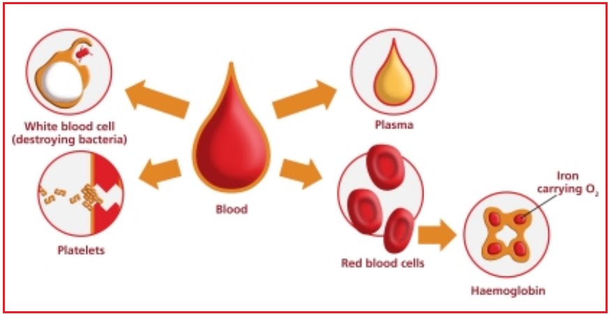
Elemen-elemen darah dibagi menjadi 3, yaitu eritrosit, leukosit dan trombosit.

Eritrosit
  • Bentuknya seperti cakram atau bikonkaf
  • Tidak memiliki inti
  • Tidak dapat bergerak
  • Banyaknya kira-kira 5 juta dalam 1 mm3 
  • Berwarna kuning kemerah merahan 
  • Berfungsi mengikat O2 dari paru-paru untuk diedarkan keseluruh jaringan tubuh 
  • Mengikat CO2 dari jaringan tubuh untuk dikeluarkan melalui paru-paru
  • Eritrosit di dalam tubuh dibuat di dalam sumsum tulang merah, limpa, dan hati


Leukosit
  • Mempunyai bentuk yang tidak tetap, bergerak dengan perantaraan kaki palsu, mempunyai bermacam-macam inti sel, berwarna bening.
  • Macam-macam leukosit menurut ada tidaknya granula:

Granulosit, sel leukosit yang mempunyai granula, terdiri dari
  1. Neutrofil: mempunyai inti sel protoplasma banyak bintik-bintik halus
  2. Eosinofil: bentuk sama seperti neutrofil, granula dalam sitoplasma lebih besar.
  3. Basofil: mempunyai inti yang teratur, pada protoplasma granulanya besar, banyak di sumsum merah

Agranulosit, sel leukosit yang tidak mempunyai granula.
1.  Terdiri dari, limfosit: bentuk ada yang besar dan ada yang kecil, didalam sitoplasma tidak terdapat granula
2.  Monosit: terdapat disumsum merah, inti sel bulat dan panjang.

Trombosit
  • Tidak memiliki inti, berperan dalam proses pembekuan darah, berukuran 2-5µm, hanya terdapat dapa darah mamalia, berwarna putih


Plasma darah
  • Dalam plasma darah terkandung: air 90 %, protein plasma 7 %, garam anorganik 0,9 %, senyawa organik asam amino, vitamin, hormon, lipid : 0,1 %
  • Zat-zat yang terkandung dalam plasma darah: fibrinogen, garam-garam mineral, protein darah,  zat makanan,  hormon, antibodi/antitoksin

Support web ini

BEST ARTIKEL